Astrocytes and Fungal Compounds: A Review of the Glial Support Literature
Half the Brain Is Not Neurons
— KANCA —
Neuroscience in popular culture begins with neurons and generally ends with neurons. Yet in the human brain, approximately half of all cells are not neurons. The remaining half consists of glial cells, and the largest member of this glial population is the astrocytes.
Astrocytes are called "support cells." This label is misleading: astrocytes are responsible for the brain's chemical homeostasis, clearing the synaptic cleft, maintaining the blood-brain barrier, and supplying metabolic substrates to neurons. Without them, neurons become dysfunctional within hours. In this article, we examine astrocyte biology and its position in in vitro studies conducted with mushroom components.
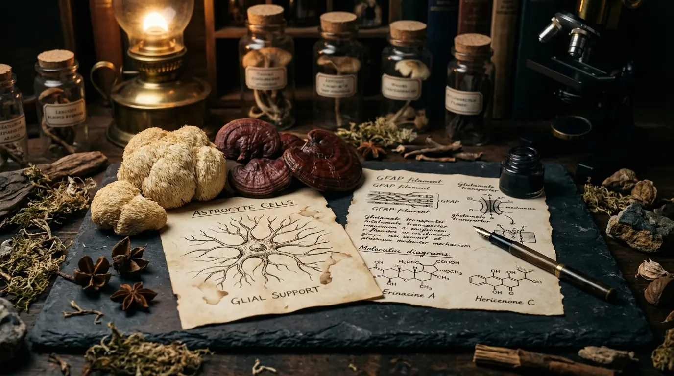
Astrocyte: The Star-Shaped Cell
The name "astrocyte" derives from its star-shaped morphology. Dozens of extensions radiate from the cell body, spreading across a wide expanse of brain tissue. These extensions reach three primary targets:
- Neuronal synapses: Astrocyte processes envelop the synaptic cleft. They rapidly clear neurotransmitters released at the synapse—particularly glutamate—and ensure signal termination.
- Blood vessels: Astrocytic end-feet cover the surface of brain capillaries. This constitutes an essential component of the blood-brain barrier.
- Other astrocytes: Astrocytes connect to one another via gap junctions. This enables the astrocyte network to operate in a synchronized manner.
This tripartite connectivity establishes the astrocyte as the brain's "structural-chemical-metabolic coordinator." A single astrocyte surrounds thousands of synapses.
Astrocyte Functions
The core functions of astrocytes include:
- Glutamate uptake: Glutamate released at synapses is taken up by astrocytes via EAAT (excitatory amino acid transporters). Glutamate is neurotoxic; prolonged elevated concentrations lead to neuronal damage. The astrocyte performs this clearance work continuously.
- Glutamine cycle: The astrocyte converts glutamate to glutamine and returns it to the neuron. The neuron converts glutamine back to glutamate. This cycle is the primary mechanism of neurotransmitter recycling in the brain.
- Lactate production: Astrocytes take up glucose, produce lactate, and deliver it to neurons. This "astrocyte-neuron lactate shuttle" hypothesis is recognized as an important model in neuronal metabolism.
- Antioxidant source: Glutathione synthesis occurs largely within astrocytes; precursor molecules are supplied to neurons.
- Ion homeostasis: Potassium released into the extracellular space during synaptic activity is taken back up by astrocytes.
This multi-faceted role constitutes the technical reality beneath the "support" label assigned to astrocytes.
Astrocyte Activation: Reactive Astrogliosis
Under conditions of brain injury, infection, or chronic stress, astrocytes become "reactive." This state involves:
- Increased GFAP (glial fibrillary acidic protein) expression.
- Cellular morphology enlarges; processes thicken.
- The cytokine profile shifts.
- Glutamate uptake capacity is affected (it may increase or decrease).
Reactive astrogliosis is not a single phenotype; in some contexts it exhibits a protective profile, and in others a neurotoxic one. This phenotypic divergence is a significant research area in modern neuroscience (a distinction recognized as "A1" versus "A2" reactive astrocyte phenotypes exists).
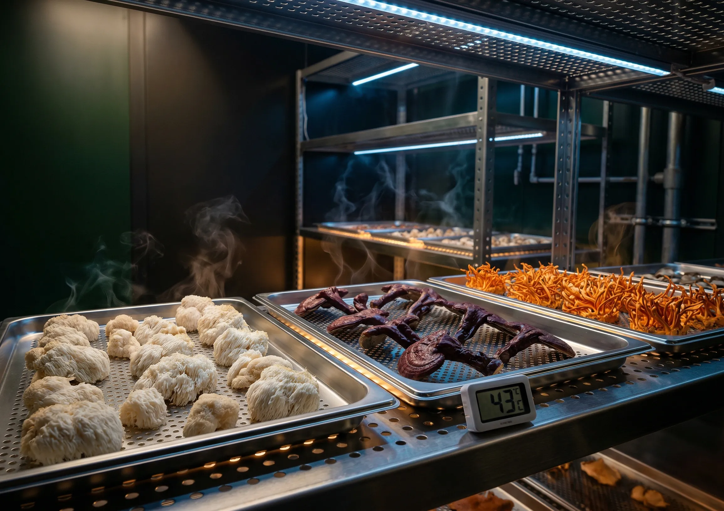
Mushroom Components and Astrocyte Literature
The in vitro literature examining mushroom components and astrocytes is limited. Study themes include:
- Lion's Mane components: Hericenone and erinacine derivatives have been studied predominantly in neuronal cell lines; however, some studies have reported NGF and BDNF profile observations in primary astrocyte cultures. This literature constitutes an in vitro research domain.
- Polysaccharide fractions: The profile of mushroom β-glucans affecting inflammatory response parameters in astrocyte culture models has been examined in a limited number of studies.
- Triterpene modulation: Studies on the inflammatory response profile of Reishi triterpenes in neuron-astrocyte co-culture models are small in scale.
This literature exists at the in vitro and animal model level. No clinical database establishes a direct pharmacological link between mushroom components and glial function.
Methodological Notes
When interpreting astrocyte in vitro studies:
- Primary culture vs. cell line: Primary astrocyte cultures approximate in vivo astrocytes. Lines such as C6 and U87 are tumor-derived astrocyte-like cells; they do not substitute for primary astrocytes.
- Purity: Primary astrocyte cultures frequently contain microglial contamination. Results obtained without verified purity may be misleading.
- Culture medium serum: Serum-containing culture conditions drive astrocytes toward a reactive phenotype. Serum-free defined media yield a more "resting" astrocyte phenotype.
- Regional heterogeneity: Astrocytes are not homogeneous across the brain; cortical, hippocampal, and cerebellar astrocytes exhibit distinct profiles. A single culture does not represent all astrocytes.
Related Readings
- What Is NGF — The biology of nerve growth factor.
- What Is Erinacin — The diterpene profile of Lion's Mane mycelium.
- Hericium Erinaceus Biological Legacy — Lion's Mane species encyclopedia.
This content is for informational purposes only and does not constitute medical advice. Consult your physician before making any health decisions. Functional mushrooms are not drugs and cannot be used to treat diseases.
Version: 1.0 | Last updated: 27 April 2026 | Sources reviewed: 16+ | Method: Editorial Policy | References: Bibliography